WCM-Q researchers use pioneering eye exam to identify nerve damage in long COVID patients
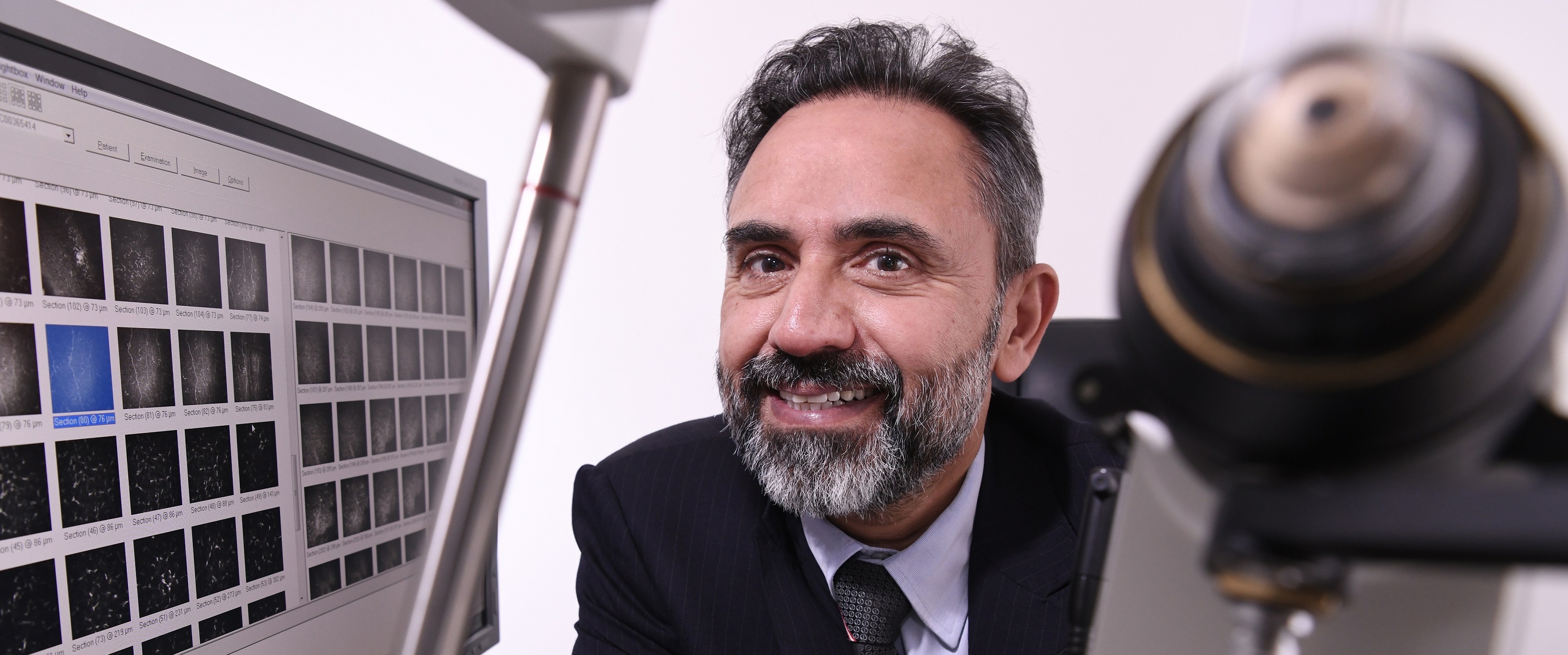 An eye examination pioneered by Dr. Rayaz Malik of Weill Cornell Medicine-Qatar can be used to detect nerve damage in long COVID patients.
Researchers at Weill Cornell Medicine-Qatar (WCM-Q) have shown that a specialized eye examination can be used to identify underlying nerve damage in patients diagnosed with long COVID.
The examination technique – called corneal confocal microscopy (CCM) – could be used as a diagnostic tool to confirm suspected cases of long COVID, say the researchers.
CCM is a non-invasive eye examination which enables real-time imaging of the corneal nerve fibers, damage to which provides a tell-tale sign of a range of neurodegenerative conditions, including diabetic neuropathy, Parkinson’s disease, multiple sclerosis, dementia – and now, long COVID. The use of CCM as a diagnostic tool for these conditions (and a number of others) was pioneered by Dr. Rayaz Malik, professor of medicine and assistant dean for clinical investigations at WCM-Q.

Upon the emergence of long COVID, Dr. Malik and colleagues identified the potential for CCM to be used to investigate the characteristics of the disease, which affects at least 10 percent of people who have recovered from acute COVID-19 infection. In a collaborative study with Dr Gulfidan Bitirgen from Necmettin Erbakan University in Konya, Turkey, they assessed for the presence of long COVID using the long COVID questionnaire of the National Institute for Health and Care Excellence (NICE) for England and performed CCM examinations on 40 patients who had recovered from COVID-19 three to four months previously.
Long COVID has been defined by the National Institute for Health and Care Excellence (NICE) in England as ‘signs and symptoms that develop during or following an infection consistent with COVID-19 and which continue for more than 4 weeks and are not explained by an alternative diagnosis’. The main neurologic symptoms of long COVID include headache, tingling and numbness, neuropathic pain, a loss or change in the patient’s senses of taste and smell, and ‘brain fog’.
Dr. Malik, who is also a practicing consultant physician, said: “The predominance of neurologic symptoms in people with long COVID prompted us to investigate whether corneal confocal microscopy could be used to objectively identify nerve damage in patients with the disease. We are the first group in the world to report a very strong association between nerve damage observed using CCM and long COVID. Although, the majority of people had mild COVID, patients with more severe disease had evidence of greater corneal nerve damage, suggesting that the severity of nerve damage may be related to the severity of disease at presentation.”
Crucially, nerve damage observed in the corneas using CCM can be reliably used as an indicator of nerve damage in other parts of the body. CCM’s value as a diagnostic tool is increased by a number of important factors: the test takes only a few minutes, is completely non-invasive, causes almost no discomfort for patients, utilizes existing and widely available ophthalmic equipment, and can be done in the clinic.
Dr. Malik added: “We believe CCM has the potential to serve as an extremely valuable tool to be used by physicians to help diagnose and assess the evolution of long COVID and to determine the severity in individual cases. The identification of underlying nerve damage also allows us to think about this condition as a neurodegenerative disease, which may be amenable to treatment.”
The study, titled ‘Corneal confocal microscopy identifies corneal nerve fibre loss and increased dendritic cells in patients with long COVID’, has been published in the British Journal of Ophthalmology.
The study can be read in full here.