WCM-Q scientist builds world’s smallest drill
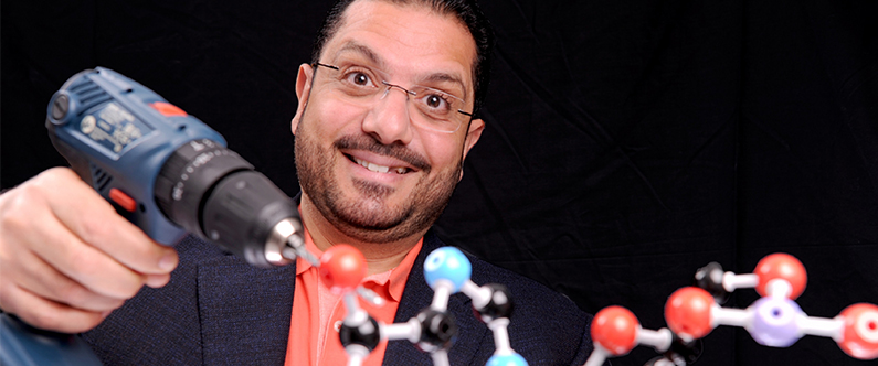 Dr. Mohammad Yousef illustrates the concept of the nano-drill he and fellow scientists have engineered from a section of protein.
A scientist at Weill Cornell Medicine-Qatar (WCM-Q) has created the world’s smallest drill – 50,000 times smaller than the breadth of a human hair.
The drill, which measures just two nanometers long, has been created and analyzed by Dr. Mohammad Yousef, associate professor of physics at WCM-Q, in collaboration with an international team of scientists from the University of Oregon, UT Southwestern Medical Center, the Max Planck Institute of Developmental Biology and Southern Illinois University Edwardsville.
The product of a decade of research, the nano-drill has been engineered from a section of protein and may have potential applications in the delivery of drugs to targeted areas within the human body.
The research team behind the drill was initially concerned with proteins and why they are structured in the way they are. But in the course of that work they discovered the potential to re-purpose a protein for alternative uses. Using a protein that in nature eats up bacterial cell walls, the team engineered a section of it to act in a different way.
Dr. Yousef, who is the lead author of the nano-drill research paper, said: “The protein’s structure had a “spring” at the surface which made it an ideal candidate for this work. We saw that a section of the spring tightens and has its motion limited when it is exposed to a chemical called guanidinium. The guanidinium essentially anchors part of the spring. When the guanidinium is removed, the spring then moves to the left. When guanidinium was added once again, the anchor was re-established and the spring moved to the right. The motion is the same that one would see in a hammer drill.”
But although the team knew that the movement occurred and had static images of the spring in different locations, they were unaware of the movement itself.
Dr. Yousef explained: “We took static pictures of the ‘on’ and ‘off’ states but we didn’t know what was happening in between and that is essential to the design if we want to use the drill in applications; we need to know exactly how the spring moves between the two locations. We need a movie, not just a static picture. The entire drill might disassemble, which we don’t want.”
The team turned to super-computing to help solve the problem and has now seen the first few frames of movement.
“We now have the first glimpse of the movie,” said Dr. Yousef. “We know the first shot, we know the last shot and now we have some of the film itself. The movement itself is very significant as it spans the entire structure of the protein, so there are undoubtedly applications out there. I envisage that it could be used for minimally-invasive procedures and personalized medicine, and it could be used to target individual cells with drugs, it could be used as a biosensor as well.”
Dr. Yousef and the research team will now continue to work on viewing the entire ‘film’ of the drill in action.
The research can be read online here.