
Grand Rounds explores novel diagnostic test for diabetic neuropathy
January, 2015
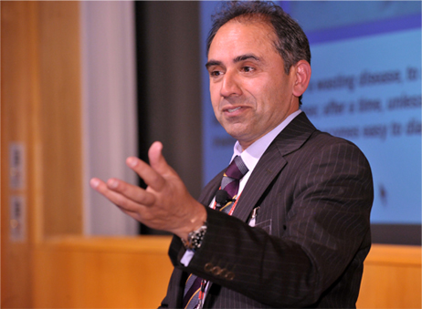
Dr. Malik told the audience that diabetic neuropathy affects 50 percent of
people with diabetes.
Pioneering research into early diagnosis and prevention of nerve damage caused by diabetes was discussed at the third installment of WCMC-Qs Grand Rounds.
Guest speaker Dr. Rayaz Malik, professor of medicine at WCMC-Q, explained that early signs of diabetic neuropathy caused by high blood sugar levels can be detected by examining the nerve fibers in the eye using a technique called corneal confocal microscopy.
The Grand Rounds, developed by WCMC-Qs Division of Continuing Professional Development, features presentations by expert speakers that aim to engage healthcare professionals in the community to enhance their knowledge of the latest developments in medical technology, research and best practice.
Dr. Benjamin Shykind, assistant professor of cell and developmental biology at WCMC-Q, introduced Dr. Malik as one of the worlds foremost authorities on diabetic neuropathy and a Fellow of the Royal College of Physicians in the United Kingdom.
Addressing an audience of fellow healthcare professionals at WCMC-Q, Dr. Malik said: Diabetic neuropathy affects about 50 percent of diabetes sufferers and causes many health problems, most typically loss of sensation in the lower limbs and feet. This can be particularly serious as it can lead to ulceration with chronic infection and subsequent amputation of the lower limbs, hence the need for early diagnosis. However, diagnosis of diabetic neuropathy poses several problems because traditionally advocated tests such as physical examinations do not return consistent results, and nerve conduction studies are not good at detecting early-stage neuropathy. Taking a skin biopsy is fairly effective but has the problem of being somewhat invasive.
“The great thing about corneal confocal microscopy is that the only slightly uncomfortable part of the examination is applying a mild local anesthetic to the patients eye using eye drops. Most importantly, it allows us to detect neuropathy at a very, very early stage.”
Corneal confocal microscopy is a relatively new technology that allows the structures of the cornea to be viewed with high magnification and very high resolution. The cornea is an attractive target for investigating nerve damage because it has the highest density of nerve fibers of any tissue in the body. These factors mean that the new diagnostic tool is so effective that it can even detect neuropathy in pre-diabetic patients those who have blood glucose levels that are higher than normal but not high enough for them to be classified as fully diabetic. Furthermore, it has applications to detect nerve damage in a range of other peripheral neuropathies, including inherited neuropathies, and drug induced toxic neuropathies, particularly chemotherapy induced neuropathy. There are also emerging data that it may even detect abnormalities in diseases of the brain such as multiple sclerosis and dementia.
Dr. Thurayya Arayssi, WCMC-Qs associate professor of medicine and associate dean for continuing professional development, said: “Dr. Maliks groundbreaking research has the potential to bring about widespread change in the way physicians diagnose diabetic neuropathy, and to allow for far earlier diagnosis than is currently the case, with obvious benefits for diabetes sufferers.
“Grand Rounds exists to share exciting and useful knowledge such as this with the healthcare community in Qatar and the wider region, for the benefit of all healthcare providers and their patients.”