
Pixelated brain comes to WCMCQ
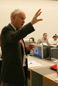
As part of the Medicine,
Patients and Society II
course, Dr. Brooks gives a
lecture on the brain and
mind to students at WCMC-Q
during his visit to Qatar.
Technological delivery of the curriculum at WCMC-Q took another step forward this semester with the arrival of the "pixelated brain" as a feature of the Brain and Mind section of the Medicine, Patients and Society II course for second year medical students.
Dr. Dana Brooks, professor of neuroanatomy in neurology and neuroscience at Weill Cornell Medical College in New York, visited Doha to deliver the sessions.
The pixelated brain is a computer program developed by Dr. Brooks for medical students in New York and Qatar which allows them to explore the brain on their own. Dr. Brooks has installed over 2000 images into the system using Dreamweaver, a design program that offers views of the brain and specific sections as well as diagrams.
Images are woven together with text to create 14 modules, each dedicated to a specific region of the brain or brain function. Each module forms the basis for a 2-hour laboratory session with an instructor who wanders through the classroom giving one-on-one help to students. A discussion then takes place, which involves a clinical problem resulting from damage to the brain pathway or region under study.
While the pixelated brain is primarily a computer program used to present new information, it contains other features. There are self-quizzes that students can use to check their own understanding of the material. There is also a set of flash cards that can be used to organize key facts.
Dr. Brooks said: "The brain is like a busy city. It is alive with activity and normally everything runs very smoothly yet the complexity of what keeps it going is hidden from view. This valuable learning tool helps the doctor to picture the brain in three dimensions, view all the sections and identify where they are, what they do and how they are connected to each other.
"Students have traditionally studied brain anatomy through viewing sections of brain tissue on glass slides. For a 100 years of more, medical students have learned neuroanatomy by sitting in a lecture room looking at projections on a screen. Studies have shown the best way for students to learn new material of this sort is for them to be actively involved in digging this information out.
"As with learning about a city, the best way is by actually going and exploring it. The experience is an essential part of the learning process and this is what this program encourages."
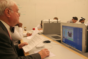
Dr. Dana Brooks takes a virtual tour of the brain
using his pixelated brain computer program.
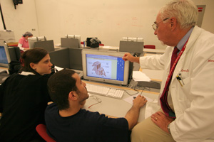
Associate professor of neurology and neuroscience
and course co-director for the second year pre-
medical Brain and Mind course, Dr. Leopold J. Streletz
(right), helps students Amila Husic and Osama Al
Saied understand a clinical problem resulting from
damage to the brain pathway.
Krista Dobinson, Assistant Editor/Writer